How do I know if I need Coolsculpting?

How do I know if I need Coolsculpting®?
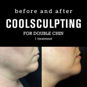
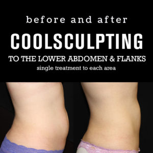
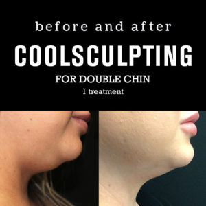
If you pinch the skin in the area you are interested in treating and are able to raise 1 to 2 inches of excess skin (fat), then you will benefit from Coolsculpting®. Coolsculpting® is designed to remove isolated pockets of fat that persist, even with diet and exercise.

The Coolsculpting® treatment works best for people whose BMI is normal, or less than 25. We recommend at least two treatments per area for best results. If your BMI is between 25 and 30, we suggest 2 to 3 treatments. We don’t recommend Coolsculpting® for persons whose BMI is greater than 30 because there will be too much fat in the target area to notice a difference after treatment. You may be a candidate for Liposuction instead. The Coolsculpting® procedure will not reduce your weight or treat obesity. Calculate your BMI.

People who have a disease that is made worse by cold or have hernias in the area to be treated should not have Coolsculpting®. People with a large amount of fat in the target area will not notice an improvement after treatment.

How does CoolSculpting® work?
The first step is to wet the skin with a moist gel pad. Then the applicator is placed in contact with the skin. When the suction is turned on you will feel pressure as the skin and fat are drawn into the applicator for freezing. When the skin is released you will feel slight discomfort for 2 to 5 minutes until the freezing takes effect and numbs the skin.

What areas can be treated with CoolSculpting®?
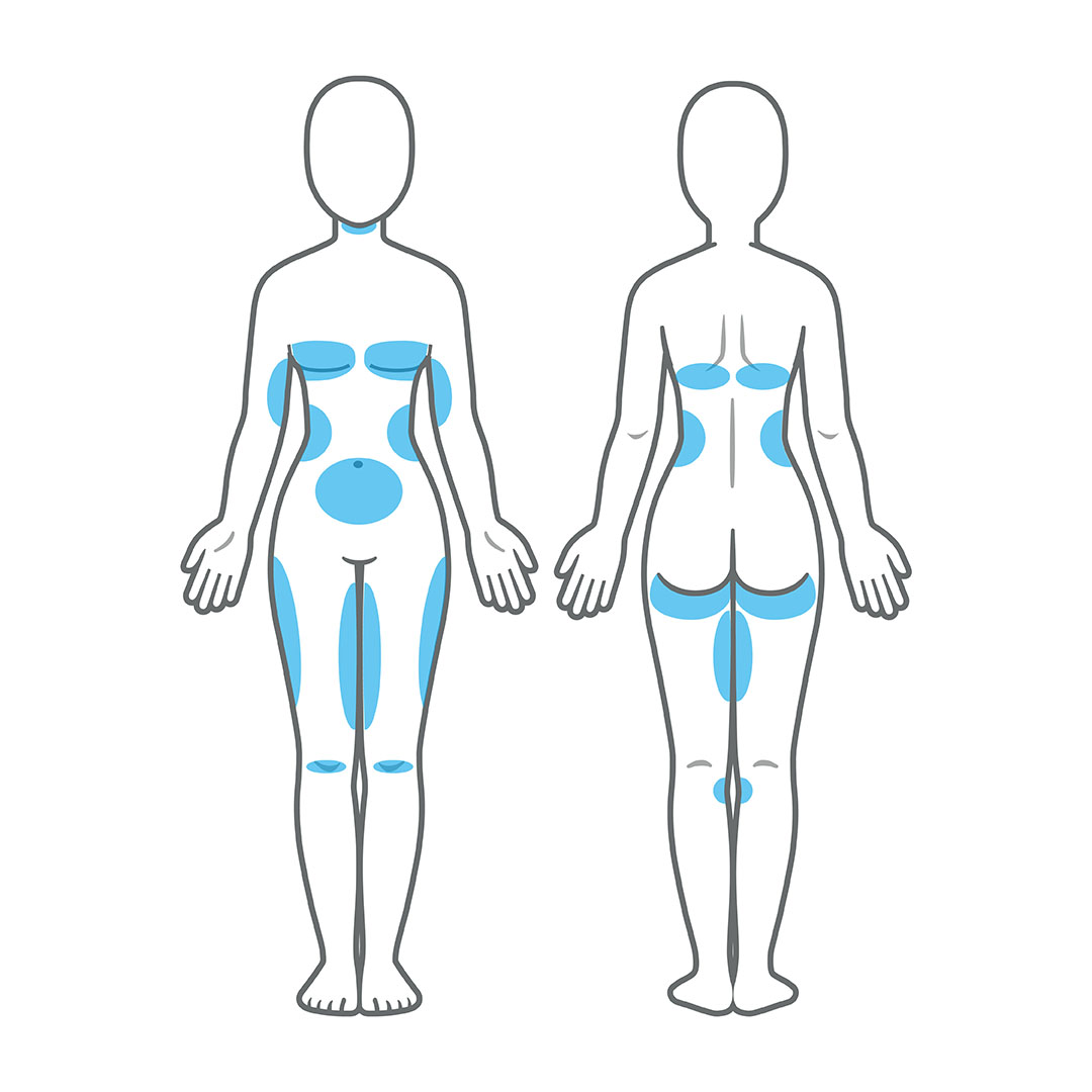
CoolSculpting® technology is used to sculpt the upper and lower abdomen, love handles, inner and outer thighs, back fat, “man boobs”, and upper arms.

CoolSculpting Areas - Lakeview Dermatology Chicago

Lakeview Dermatology Hyde Park Location

COMING SOON!
We are thrilled to announce that Lakeview Dermatology’s 4th location is coming to Hyde Park, Chicago, IL in March 2019!

Location: 1304 East 47th St, Ste 200 Chicago, IL 60653
Lakeview Dermatology Hyde Park Location

Meet Dr. Rosen

DrRosen

Hello all! This is Dr. David Rosen, the vein expert at Lakeview Dermatology. March means many things to many people: “march madness” with college basketball tournaments; spring break getaways; getting tax documents together…and you might not have known about this one…it is also “vein season”.

Vein season is the time of year when patients think about treating the varicose and spider veins that have bothered them for years. Many patients look to the summer months ahead…”shorts season” and wonder if it is possible to improve the feel and look of their legs and be able to get back into shorts and skirts more comfortably.
Vein Season
It’s absolutely possible…but it is a process.

That process begins with a consultation with me. During the hour-long visit, I get to learn about:
• Your medical history
• Any questions/concerns you may have regarding varicose and spider veins. I also spend a great deal of time doing what I love: teaching patients about vein disease!
• Your treatment goals.

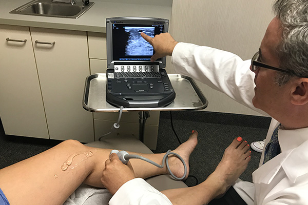
If appropriate, I am able to perform a painless and very informative ultrasound exam to help discover any underlying sources for symptoms, varicose veins and spider veins. Once the ultrasound is completed, we can tailor a treatment plan to suit your specific needs. All treatments are performed in our Lakeview Dermatology offices: you can drive yourself to/from any treatment visit. There is no down time and few restrictions after treatments: most patients resume their normal routines immediately.

UltrasoundExam

Many patients are also pleasantly surprised to learn that their consultation visit, ultrasound and many treatments may be covered by insurance, including Medicare! However, it is important to understand that coverage for treatments, when available, may require weeks to months to achieve.

That’s why it is so important to schedule your initial consultation visit with me as soon as possible. Let’s make this “vein season” the one that begins your journey towards “shorts season” and a life with happier, healthier legs!

Call 773-281-9200 now to schedule your appointment with me. See you soon!

Study shows that Nicotinamide may help prevent Malignant Melanoma

25254389 - nicotinamide adenine dinucleotide (nad+) coenzyme molecule. important coenzyme in many redox reactions. atoms are represented as spheres with conventional color coding: hydrogen (white), carbon (grey), nitrogen (blue), oxygen (red), phosphorus (orange).

25254389 – nicotinamide adenine dinucleotide (nad+) coenzyme molecule. important coenzyme in many redox reactions. atoms are represented as spheres with conventional color coding: hydrogen (white), carbon (grey), nitrogen (blue), oxygen (red), phosphorus (orange).

A new study from Australia suggests that Nicotinamide may help prevent Malignant Melanoma.
In a review published in the journal Photodermatology, Photoimmunology& Photomedicine, Professor Gary Halliday of the University of Sydney, Australia and associates, presented their findings.
It is well known that Ultraviolet Radiation damages the DNA in the cell nucleus. Professor Halliday and his team lists studies showing that Nicotinamide can boost DNA repair following UV exposure.
In addition, Nicotinamide can also reduce immunosuppression and inflammation,  both of which are factors in cancer progression.
Dr Halliday and his team have demonstrated in a recent study that Nicotinamide 500 mg daily reduced the incidence of non melanoma skin cancer by 23%. Dr Halliday and his team state that larger studies are now warranted to determine the efficacy of Nicotinamide for prevention of Malignant Melanoma
In the meanwhile, using sunscreen, seeking the shade and wearing sunglasses, sun protective hats and clothing remain the primary skin cancer prevention methods.

Sunscreen smarts

Sunscreen - 1200x628

March 21, 2017

By Bob Kronemyer – Modern Medicine Network

Sunscreen - 1200x628

Do dermatologists believe that high-SPF sunscreens provide an additional margin of safety? How safe are sunscreens that contain oxybenzone? These answers and more appear in the results of a recent survey in the January edition of JAMA Dermatology.

“Multiple professional organizations, including the American Academy of Dermatology, as well as dermatologists themselves, have recommended patient counseling regarding the use of sunscreen,” write the authors from the University of Miami Miller School of Medicine in Florida. “However, there have been other conflicting messages about sunscreen (sometimes without scientific support) that have led to confusion by the public.”

Case in point, of the 156 dermatologists included in the outcome analyses, 99% believe that their patients generally do not apply sufficient sunscreen.

As for patient counseling, 97% of dermatologists indicate they are comfortable recommending sunscreens with a sun protection factor (SPF) of at least 50, and 83.3% believe that high-SPF sunscreens provide an additional margin of safety.

When recommending sunscreen, dermatologists consider several factors, including SPF level (99%), broad-spectrum protection (96%), cosmetic elegance and/or feel (71%) and photostability (42%).

As for the doctors themselves, they have good intentions, but don’t always follow through. In their own outdoor use, 100% of dermatologists select a sunscreen with an SPF of at least 30, but only 76% admit to routinely applying sunscreen more than half the time.

Other points that dermatologists agree on include:

  • Routine use of sunscreen helps lower the risk of skin cancer (97%)
  • Sunscreen reduces subsequent photoaging (100%)
  • FDA-approved sunscreens currently available in the U.S. are safe (96%)
  • Sunscreens containing oxybenzone or retinyl palmitate are safe (91% and 87%, respectively)

In addition to counseling patients, 99% of respondents also recommend that their family and friends use sunscreen.

There were no significant differences in responses based on geographic location or number of years in practice.

Global perspecitves on cosmetic fillers

Haasan I. Galadari, M.D.

With so many new and exciting filler products in the U.S. pipeline, American dermatologists and their patients are in for a treat, according to Hassan I. Galadari, M.D., who presented “Up and Coming Fillers: Experience from Across the Atlantic,” on March 4 at the 2017 American Academy of Dermatology (AAD) Annual Meeting in Orlando, Fla., March 3 through 7.

Dr. Galadari, an American board-certified dermatologist who practices in Dubai, says there are more than 80 companies that produce hyaluronic acid (HA) fillers worldwide.

“…though not all will be approved in the U.S., the larger companies–those who push the envelope in the areas of research and development–will be introducing their products to the U.S. very soon,” Dr. Galadari tells Dermatology Times.

A trend among filler companies is to focus on adding products that cater to certain parts of the face, he says.

“In the U.S., doctors are made to dilute their fillers in order for them to safely use them in certain parts of the face–say the tear troughs under the eyes, for example. That will change with the introduction of newer softer products that have been approved for those indications,” Dr. Galadari says.

Recent U.S. approvals for Restylane’s Refyne and Defyne (Galderma) fillers occurred long after the rest of the world began using those fillers. The newest Galderma HA fillers in the U.S. have been in use in other parts of the world since 2010, as part of Galderma’s Emervel line.

HA fillers are not the only potential new kids on the block. Non HA fillers will be entering the U.S. market soon. While not much has changed with poly-L-lactic acid (PLLA) and calcium hydroxylapatite (CaHA) formulations, a filler containing polycaprolactone (PCL) is in the pipeline to be approved in the U.S., according to Dr. Galadari.

“PCL is a bio-stimulatory filler that comes in four different formulations of different longevities, without affecting its viscosity and no change in extrusion forces. The longest in the range, which can last up to four years, may be used for those with structural anomalies or defects, such as HIV lipoatrophy and even acne scars,” he says.

Avoiding, addressing potential complications

Filler use is not without complications. The good news is there are ways to address and avoid them.

Swelling is one of the most common adverse filler-related effects. Different fillers—even those in the same category, such as HA—behave differently, according to Dr. Galadari.

“Depending on the concentration of short and long chain HAs in the product, the degree of swelling varies. Swelling is transient, but there are some products where the swelling can last more than two weeks, and, for patients seeking fast and immediate beautification for an event, [that might not be acceptable],” he says.

Bruising can result, especially when treatment involves many injection sites.

“Needles, potentially, can statistically cause more bruising to happen as compared to a cannula. That’s why the use of cannulas have risen. Cannulas are great tools, but their limitation may lie in the fact that they can only inject the deeper plane and not the dermis. The superficial lines need to be injected with needles,” Dr. Galadari says.

Lumps can occur with use of any product. And when they do with HA fillers, injectors can simply dissolve them by injecting hyaluronidase.

“The same cannot be said with non HA fillers, which may require the injection of saline to help soften the lump, but not essentially dissolve it. Granulomas are distinct lumps that have to be proven histopathologically,” he says.

Biofilm can occur with any product but are common with HA fillers. The only way to remove biofilm is with complete removal of the product and a course of antibiotics, according to Dr. Galadari.

“Intralesional steroid use has fallen out of favor and is not as common as in the past, given that [the steroids] may cause more harm than good in terms of causing atrophy to the surrounding tissue,” he says.

Neurovascular compromise, while not the most common adverse event, is the most alarming. Necrosis can ensue if it is not correctly identified. If that happens, the effect is usually long lasting scarring. It is important to be able to identify this as quickly as possible, according to Dr. Galadari.

“Patients may present in two different ways. The first is immediate blanching of the area where the filler is being injected. This identifies an embolism, an intravascular injection of the filler. If this occurs, then immediate cessation of injection should be done, followed by flushing of the filler with a high concentration of hyaluronidase for both HA and non HA fillers. Manual massage is important as this recruits surrounding blood vessels to the area. Warm compresses may also be applied. [Nitroglycerine] paste may also be used. Patients should be injected repeatedly with hyaluronidase every 30 to 60 minutes before leaving the clinic to ensure that circulation has been reestablished in the area. Aspirin and sildenafil have also been prescribed to be used at home as an antiplatelet and vasodilator respectively,” he says. “The other feature of impending necrosis can occur when at home. Patients usually complain of severe pain and tenderness within the first 24 hours. If patients do so, the doctor should immediately ask them to come to the clinic and the steps delineated above should be performed.”

On the horizon

Teoxane’s Teosyal Resilient Hyaluronic Acid (RHA) line will introduce one of the newer fillers to the U.S. market, according to Dr. Galadari.

“This will be distributed by Alphaeon in the US. The filler will be approved by the end 2017 or beginning of 2018,” he says. “One of the advantages of this filler is its ability to stretch in areas of dynamism, especially the mouth, but without compromising its lifting effect. The filler holds an advantage of becoming integrated to the surrounding tissue and is extremely forgiving, in the unlikely event of when a lump occurs. Preliminary work in the lips and perioral lines are very promising.”

Other newcomers to the U.S. filler market, according to Dr. Galadari, include additions to the Merz Aesthetics Belotero line, including a volumizing HA. Galderma and Allergan will also continue to introduce fillers that have been available in Europe and the rest of the world, he says.

Dr. Galadari offers these three best practices for filler use:

Choose the right patient. If you don’t have the right patient and decide to inject anyway, you will have an unsatisfied patient. Ask patients specific questions to understand their expectations and to determine whether filler injections are right for them.

Choose the right filler for the right area. It’s extremely important for doctors to understand filler properties, both physiologically and histologically. A dermatopathologist showcased the importance of this point during Dr. Galadari’s AAD session.

Some examples: If a filler with high elastic modulus, or G’, is injected in the lips or tear troughs, the filler will have a tendency to create highly undesirable lumps. The same thing can be said if the injector uses fillers with a lower G’ in the cheeks. He or she will need a huge volume to get a lifting effect, which can be quite costly for the patient. That being said, biostimulatory fillers should be avoided in general in areas of the lips and tear troughs. With the upcoming introduction of softer HA fillers , areas which were considered taboo, such as the glabellar complex, where many reported cases of necrosis have occurred in the past,  may now safely be injected provided that anatomic knowledge of the area is adequate.

“Currently, there are low-G’ HA fillers that can be safely injected in that area provided a ‘correct’ approach is undertaken to avoid pitfalls,” he says. “With the newer fillers being approved, we can now safely inject the correct filler for those anatomical spots.”

Choose the right injection technique. Recent adverse events (even blindness) resulting for cosmetic filler treatments draw attention to what can happen when injectors use inappropriate injection technique and have limited understanding of the anatomy.

Disclosure: Dr. Galadari is a speaker, faculty member or investigator for Sinclair Pharma (previously Aqtis Medical), Merz Aesthetics and TEOXANE Laboratories.

The Skinny on fat reduction, cellulite devices

Coolsculpting January 18, 2017

By Lisette Hilton – Modern Medicine Network

Devices aimed at reducing fat and ridding patients of cellulite’s unsightly bumps look magical in promotional ads, but how do dermatologists look beyond to choose the best technological arsenal for treating their patients? We asked for unplugged advice from Chapel Hill, N.C., dermatologist Sue Ellen Cox, M.D., who presented on fat reduction and cellulite devices in November at the 2016 American Society for Dermatologic Surgery (ASDS) annual meeting in New Orleans.

The reveal

There are plenty of noninvasive fat reduction devices. Nuances help differentiate them.

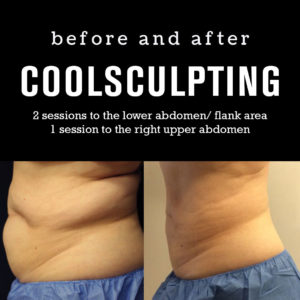
Dr. Cox’s noninvasive fat-reduction device of choice and the one that she has been using for about five years is CoolSculpting (Zeltiq).

The ultrasonic destruction of fat cells with UltraShape is another option. Dr. Cox did the clinical trials for UltraShape but doesn’t own the device.

“The company typically recommends three treatments to get the result that is expected or anticipated by the patient,” Dr. Cox says. “What I was seeing during the clinical trials was that it may have not been quite as predictable as what I see with the CoolSculpting, which is typically just one or two treatments for a very defined 20 percent to 25 percent reduction of fat. I can also tell you in terms of studies out there, CoolSculpting for noninvasive fat reduction really does have the most.”

Other options: Vanquish (BTL Aesthetics), a radiofrequency device for fat reduction, and the other ultrasonic device Liposonix (Valeant).

“I had the Liposonix done on my lower abdomen once and didn’t even finish the procedure it was so uncomfortable. And I was not overly impressed with the results that I was seeing in the clinical trials,” Dr. Cox says. “I had the CoolSculpting done on my lower abdomen once and upper abdomen twice with no discomfort.”

The new device on the block is SculpSure (Cynosure), which is a noninvasive laser.

“It’s a 1060 nm wavelength laser that is attracted to fat as its chromophore,” Dr. Cox says.

Dr. Cox says she thinks all these devices are good, but some work better than others.

“Some of them require more sessions, like the UltraShape or Vanquish. Even the SculpSure requires more than one treatment. When CoolSculpting first came out, they talked about it being a one and done thing, but over the years have adjusted expectations by saying there’s a ‘treatment to transformation,’ meaning that you treat an area and you’re going to get 20 to 25 percent reduction in fat. But you may want to treat it again, to get another 20 to 25 percent reduction, or work areas that are adjacent to the areas that you’ve already worked on, so you’re truly sculpting these patients,” Dr. Cox says.

Liposuction still hard to beat (track record-wise)

Though she says these numbers might have since changed, Dr. Cox did a PubMed data search in October 2015 and found there were 50 peer-reviewed articles on CoolSculpting; 37 for Liposonix; 18 for UltraShape; 16 for Vanquish; and eight for Kybella. Cynosure’s website lists three paper presented at American Society for Laser Medicine and Surgery meetings but no published articles in the peer-reviewed literature.

“But if we compare published studies to liposuction, liposuction has over 1,190 peer-reviewed articles. So, liposuction still may be the gold standard, even though that’s not what patients appear to be as interested in because it’s invasive and there’s downtime,” Dr. Cox says.

While liposuction’s appeal might wane in light of new technologies, patients who do opt for the old standby fat removal approach are generally as happy as or happier than those who have noninvasive options.

“If you look at RealSelf, and I pulled the data from 2015 and 2016, it’s across the board in terms of people saying they’re really happy with [liposuction],” Dr. Cox says. “Typically, liposuction has one of the highest ‘Worth It’ ratings and number of reviews,” she says.

For example, at last glance (December 5, 2016), liposuction had 4,561 RealSelf reviews and a 91 percent Worth It rating; UltraShape had 115 reviews and an 87 percent Worth It rating. For CoolSculpting, there were 2,630 reviews and an 83 percent Worth It rating. And Kybella had 279 reviews and 87 percent Worth It rating.

“But I do think that patients are happy with the noninvasive technology. It is pretty easy with the CoolSculpting. The nice thing is they’ve updated the device, so there is a 35-minute cycle time, as opposed to an hour. From the standpoints of patient ease and satisfaction, it’s great,” Dr. Cox says. “In terms of patients being able to return to work with any of these noninvasive devices, it’s pretty immediate. The next day, they can go back to exercising; they can go back to work. There’s really no downtime, with the exception of maybe some temporary numbness or tingling sensation.”

But patients might need multiple sessions with noninvasive devices, whereas, it’s one treatment and done with liposuction, Dr. Cox says.

“You can do several areas at once and have one downtime, one procedure, one expense,” she says.

Dr. Cox says her colleagues that offer fat reduction and removal should keep in mind that the noninvasive are becoming more and more of a commodity offered at salons, medi spas and more.

“There’s a spot near me that was only doing laser hair removal until about six months ago. Now they’re doing CoolSculpting. That’s an issue,” she says.

Making choices

Dr. Cox says colleagues should learn before they purchase or rent one of these devices.

“Definitely look at the peer-reviewed data,” she says. “That’s why I chose CoolSculpting. There is a lot of data to show it is predictable—that it results in 20 to 25 percent reduction in fat every time you’re hooked up to the device. There was recent information where there was 30 percent reduction with UltraShape, and maybe that’s 30 percent because you have to do it three times. You have to look at all this information critically,” she says.

Dermatologists who don’t hop on the noninvasive fat reduction train might miss opportunities for new patients, Dr. Cox says.

“If you don’t have the noninvasive technology, patients aren’t as likely to be driven to your door. They’re drawn to your office by what they see online or on billboards. If you don’t have the noninvasive technology, they’re less likely to come to your office and they may be a better candidate for liposuction but you might not even get to see them,” she says.

The field for noninvasive fat reduction is burgeoning.

“The demand is so high. Patients calling the office. Who doesn’t have a bulge they’d like to get better?” Dr. Cox says.

Disclosure: Dr. Cox did clinical trials for Liposonix (Valeant Pharmaceuticals), UltraShape (Syneron Candela) and Kybella (deoxycholic acid, Allergan). She is a consultant for Merz.

Zika – What You Need To Know

July 25, 2016
By Maureen S Hamel MD, Brenna L Hughes MD MSc – Modern Medicine Network

60980924 - doctor hand touching zika virus sign on virtual screen. medical concept

Zika virus was first identified from the Rhesus monkey in 1947 in the Zika Forest, Uganda. The virus was subsequently isolated from humans 5 years later. Since the 1950s 3 major outbreaks have been reported: 2007 in the Yap islands of Micronesia; 2013 in French Polynesia; and most recently in 2014–2015 in Brazil. Because the symptoms of Zika are similar to several other viruses such as chikungunya and dengue fever, it is likely that cases in other regions have occurred but have not been identified.

Prior to 2007, Zika was not reported outside of Africa and Asia, and since it was first reported in Brazil in May 2015, the virus has swiftly spread across South and Central America and into the Caribbean. During this time, Brazil has seen a concomitant increase in the frequency of neonatal microcephaly. These parallel findings suggested a link between Zika and birth defects—a relationship not previously identified. With the outbreak of Zika infection and its probable link to teratogenicity has come vast media attention. Zika was declared a Public Health Emergency of International Concern by the World Health Organization (WHO) in February 2016 and it is predicted that transmission to new countries and territories will continue during the upcoming months to years. In fact, in June 2016, the WHO took the momentous step of recommending that women living in Zika-endemic areas delay child-bearing.

Vector

Zika is transmitted to humans via the Aedes species of mosquitoes. These mosquitoes are also responsible for the transmission of dengue fever and chikungunya viruses. The Aedes mosquitoes are unique; unlike other species of mosquitoes, they are aggressive daytime biters. The primary species transmitting Zika to humans is the Aedes aegypti and the secondary species is the Aedes albopictus. The virus is transmitted during a blood meal. Both Aedes aegypti and Aedes albopictus have been found in the United States, primarily in the southeastern region. Aedes albopictus is the vastly more prevalent species (Figure 1).

obgyn0816_017_1

Disease features

Zika is a flavivirus, a single-stranded RNA virus. Zika virus is challenging to identify and therefore challenging to study for several reasons.

  1. First, only 1 in 5 people infected with the virus will become ill.
  2. Not only are disease symptoms usually mild, they can be identical to many other common mosquito-transmitted viruses. Because mild viral illnesses are ubiquitous in many of the regions affected with Zika, people may never realize they have been infected. In patients who do experience symptoms, these symptoms will occur between 3 and 12 days after being bitten by an infected mosquito and will last between 2 and 7 days. Symptoms include mild fever, maculopapular rash, headache, arthralgias, and myalgias. One distinctive feature of Zika is non-purulent conjunctivitis; while this complaint is also found among patients with dengue fever and chikungunya, its presentation seems to be much more common and severe in Zika cases. Zika has not been shown to cause hemorrhagic fever, and disease requiring hospitalization and/or resulting in death is rare. As in the case of other viral illnesses, once infected, individuals are protected from future infection.

Infection prevention

The most effective methods of disease prevention are to protect against mosquito bites and to reduce opportunities for vector proliferation. The Centers for Disease Control (CDC) has recommended long-sleeved shirts and long pants in Zika-infected regions both in the daytime and at dusk. Mosquito bed nets are essential if air conditioning or screens are unavailable. The CDC also recommends the application of Environmental Protection Agency (EPA)-approved repellents to skin and clothing. These products include DEET (N,N-Diethyl-meta-toluamide) and premethrin. DEET-containing products are recommended for skin application and provide the longest-lasting skin protection. Premethrin should be used on clothing and netting. Products approved by the EPA are not expected to cause adverse health effects and are safe in pregnancy. In fact, reports have suggested it may soon be possible in some states to receive Medicare/Medicaid for DEET products. Finally, vector reduction is essential for disease pre vention. The CDC has advised state and local governments to urge residents to reduce standing water by eliminating old tires, old barrels, and other vessels for standing water. An initiative has begun to treat standing water and wetlands with larvicides such as the bacterial insecticide Bti (Bacillus thurengiensis israeliensis).

Transmission

Although Zika was discovered more than 60 years ago, because adverse pregnancy or birth outcomes have never been previously reported, research into perinatal infection and perinatal transmission is limited and ongoing. Two theories of perinatal transmission exist. The first is transplacental transmission. This theory proposes that virus is transferred directly from the mother to the fetus via the placenta. Once infected, the fetus suffers neural damage as a direct result of the virus. The other theory is one of placental inflammation, which proposes that maternal viral infection creates a placental inflammatory response. This response in turn results in fetal neural damage. The former theory is similar to other documented models of viral transmission and poor outcomes and is the more widely accepted view.

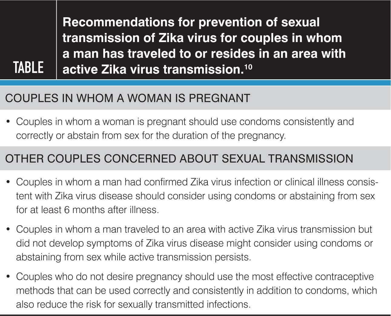
Zika can be sexually transmitted from men to their sexual partners and the possibility of male-to-female sexual transmission with subsequent fetal transmission raises concern. The first documented case of sexual transmission occurred in 2008; this was male-to-female with the sexual contact occurring a few days before the onset of symptoms in the man. With regard to the current outbreak, the first report of sexual transmission was in February 2016. However, as of March 2016, 6 cases of sexual transmission of Zika from men to their sexual partners had been confirmed in the United States. All cases of sexual transmission have been from a man to his sexual partner via vaginal, anal, or oral sex and the sexual contact has occurred before, during, or soon after resolution of symptoms consistent with Zika infection. One recent report out of New York City suggests the first case of female-to-male transmission, however it remains under investigation and it is currently unknown whether asymptomatic men can transmit virus to their sexual partners.

The duration of Zika within the male genitourinary tract is unclear and the process of viral shedding within the genitourinary system is not well understood. As such, Zika testing for the sole purpose of assessing the risk of sexual transmission is not recommended. Because of the risks of transmission from men to women and particularly the risk to the fetus during pregnancy, the CDC has set forth guidelines regarding sexual activity (see bellow)

obgyn0816_018_1

Two cases of peripartum transmission (ie, transmission at delivery or in the immediate postpartum period) from mother to infant have been reported; both cases resulted in mild disease for the mothers and neonates and no long-term morbidity. While viral particles have been detected in breast milk, breastfeeding has yet to be documented as a mode of transmission. Likewise, infection status after blood transfusion has yet to be reported; however, it is expected, as Zika was found in 2.8% of donors during the 2013 French Polynesian outbreak.

In June 2016, the NEJM published the first report of an affected fetus in the United States. Driggers et al. describe the case of a 33-year-old woman who traveled to Guatemala, Mexico, and Belize during her 11th week of pregnancy. A day after her return to the United States she developed symptoms of a viral infection including rash, mild fever, myalgia, and ocular pain. Her partner had similar symptoms. Serologic testing was positive for both IgM and IgG antibodies against Zika, consistent with Zika infection. Fetal ultrasounds after symptom resolution were negative for intracranial pathology. At 19 weeks, a detailed ultrasound demonstrated brain abnormalities such as bilateral frontal horn enlargement, upward displacement of the third ventricle, absence of the cavum septum pellucidum, and a decrease in head circumference from the 47th to the 24th percentile for gestational age. Fetal MRI confirmed abnormalities of the lateral ventricles, the third ventricle, and the corpus collosum. The patient elected for termination at 21 weeks’ gestation. Postmortem examination of the fetus confirmed the presence of Zika in the amniotic fluid, placenta, fetal brain, muscle, liver, lung, and spleen.

The research regarding perinatal Zika infection, while at times compelling, is limited. And many questions remain. The challenge for patients as well as practitioners is facing the unknown. The true incidence of Zika among pregnant women is unclear, as is the rate of vertical transmission. In turn, among fetuses to whom the virus is transmitted, the rate and range of clinical manifestations cannot be predicted. Indeed it seems there is a delay between maternal exposure, fetal transmission, and ultrasonographically detectable abnormalities, but this time course is not well understood. The case by Driggers et al. demonstrates that fetal abnormalities may not be seen for up to 9 weeks after maternal exposure.

Finally, and perhaps most distressing to expectant mothers, is the unknown prognosis for infants born with Zika infection. Using data from the current outbreak in Bahia as well the previous outbreaks in Micronesia and French Polynesia as models, Johansson and others estimate the risk of microcephaly among infants of mothers infected by Zika to be between 0.9% and 13%. While this estimate is concerning, it must be considered with caution as it is based on modeling and limited retrospective data. Likewise, microcephaly is only one of several potentially adverse outcomes. It is well established that infants with severe microcephaly from other causes will experience a range of neurologic sequelae, but it is unclear if this is true for all cases of Zika-related microcephaly. Neurologic problems can range from intellectual disability to sensory deficits to seizures; they can be mild, severe, or life-threatening. Long-term outcomes are not at all clear and will not be for years to come.

Countries where pregnant women are advised not to travel:

  • American Samoa
  • Argentina
  • Aruba
  • Bahamas
  • Barbados
  • Belize
  • Bolivia
  • Bonaire
  • Brazil
  • Cape Verde
  • Caribbean
  • Chile
  • Colombia
  • Costa Rica
  • Curacao
  • Dominican Republic
  • Ecuador
  • El Salvador
  • French Guiana
  • Guadeloupe
  • Guatemala
  • Haiti
  • Honduras
  • Jamaica
  • Martinique
  • Mexico
  • Nicaragua
  • Panama
  • Paraguay
  • Peru
  • Puerto Rico
  • Samoa
  • Saint Martin
  • Suriname
  • Tonga
  • U.S. Virgin Islands
  • Uruguay
  • Venezuela

Common pediatric disorders in skin of color

SkinColor2

August 10, 2016
By Lisette Hilton / Dermatology Times

While pediatric atopic dermatitis and acne have some similarities among skin of color and lighter-skin children, there are important differences when these common skin conditions affect darker skin types, according to Nanette Silverberg, M.D., clinical professor of dermatology and pediatrics, Icahn School of Medicine at Mount Sinai and chief, pediatric dermatology, Mount Sinai Health System.

Starting with eczema

Atopic dermatitis is the most common skin condition of childhood and affects about 25 percent of children in the U.S., according to Dr. Silverberg, who presented on the topic at The Skin of Color Seminar Series, held earlier this year in New York City.

“In particular, there have been studies that have shown atopic dermatitis is more common in children of African American descent or of Afro-Caribbean descent,” she says. “It certainly represents a very concerning issue in children of color.”

Differences in atopic dermatitis can occur in the presentation and severity among children of color.

“In somebody who is very light skinned, eczema is going to be red. But in children of color, we see much less erythema. We see much more in the way of lichenification, or thickening of the skin, and more follicular prominence. These are particularly vexing types of eczema, in that the lichenification, or lichenoid, type of dermatitis is often very thick and very itchy. And the follicular type can be quite deceptive. You don’t see redness. You don’t necessarily see thick or oozing skin, but it is incredibly itchy and it significantly affects children psychologically,” Dr. Silverberg says.

One of the major issues with treating children of color is that there are differences biologically, in terms of the basis of atopic dermatitis, according to the dermatologist.

In African American children, it has been demonstrated that there are reductions in ceramide content, and that could be the reason the skin barrier is not working as effectively as it should be. In children who are Caucasian of European descent, eczema is more associated with a filaggrin defect, she says.

“Filaggrin defects, particularly in Asian children, are somewhat different than those noted in Caucasian children, so we know there are some reasons biologically that the kids may be a little different,” Dr. Silverberg says.

As a result, dermatologists treating children of color who have eczema often need to use thicker emollients, including emollients that might have extra ceramide content or extra balanced fat content to enhance the skin barrier.

“We’re still moving forward to see whether the biologic basis of eczema affects how children respond to treatment. In atopic dermatitis, many of the kids with atopic dermatitis will manifest in early childhood with a lot of hypopigmentation or lightness of the skin. So, pigmentary alterations, which we see in kids of color, are temporary but are sometimes very noticeable and can concern parents,” Dr. Silverberg says. “But this generally resolves, and that’s something we can reassure parents about.”

Acne

Acne is common and comes with different concerns in children with skin of color.

“Whereas many of our Caucasian patients talk about the actual pimple lesions, most of our African American patients and many of our Hispanic and Asian patients will obsess over post-inflammatory pigmentary alterations after their acne clears,” Dr. Silverberg says. “So, there’s a focus in the skin of color acne patients, even in the teenagers, on specifically pigmentation issues.”

Hispanic pediatric patients tend to have the most severe acne types among children of skin of color, Dr. Silverberg says.

“We don’t see as much in the way of cystic acne in African American patients, historically and in the literature,” she says. “So, the population that we tend to focus on for more severe treatment or treatment, like isotretinoin, are usually Hispanic teenagers. It’s an important consideration because they have some tendency to have the cystic component, although you can see it in everybody, it seems to be the most concerning amongst that population in the teenage years.”

Dermatologists treating these children need to pay special attention to communicating the need for using good sun protection to enhance pigmentation returning properly. It’s also important to work with patients to develop a skincare regimen that’s effective both at clearing current lesions and preventing new lesions, so the pigmentation improves over time, according to Dr. Silverberg.

“There are some wonderful new acne guidelines that have come out recently from the American Academy of Dermatology … saying it’s clear that most patients of color will respond quite nicely to the products we have available, including topical retinoids … as well as azelaic acid, which has been demonstrated to be beneficial in improving both tone and skin lesions,” she says.

Study shows poor skin cancer survival in patients with skin of color

Dermatologist urges everyone to be aware of their risk and take steps toward prevention, detection
SCHAUMBURG, Ill. – July 28, 2016

iStock_000007711669Medium-1024x468

 Because Caucasians have a higher skin cancer risk than the general population, people with skin of color may believe that they don’t need to be concerned about this disease — but new research reveals this to be a dangerous misconception.

According to a study published online in the Journal of the American Academy of Dermatology on July 28, although melanoma incidence is higher in Caucasians, patients with skin of color are less likely to survive the disease.

“Everyone is at risk for skin cancer, regardless of race,” says board-certified dermatologist Jeremy S. Bordeaux, MD, MPH, FAAD, one of the study authors. “Patients with skin of color may believe they aren’t at risk, but that is not the case — and when they do get skin cancer, it may be especially deadly.”

Researchers at Case Western Reserve University in Cleveland utilized the National Cancer Institute’s Surveillance, Epidemiology and End Results database to study nearly 97,000 patients diagnosed with melanoma, the deadliest form of skin cancer, from 1992 to 2009. Although Caucasian patients had the highest melanoma incidence rate, they also had the best overall survival rate, followed by Hispanic patients and patients in the Asian American/Native American/Pacific Islander group.

African-American patients had the worst overall survival rate, and they were also the group most likely to be diagnosed with melanoma in its later stages, when the disease is more difficult to treat. According to the study, however, the timing of the diagnosis is not the only factor that affects this group’s survival rates, as African-American patients had the worst prognosis for every stage of melanoma.

Dr. Bordeaux says these differences in survival rates may be due to disparities in the timeliness of melanoma detection and treatment among different races; for example, patients with skin of color may not seek medical attention for irregular spots on their skin because they don’t believe these lesions pose a risk. Additionally, he says, there may be biologic differences in melanoma among patients with skin of color, resulting in more aggressive disease in these patients. More research is necessary to determine why survival rates differ among different ethnic groups, he says, but in the meantime, patients of with skin of color should be aware of their skin cancer risk.

“Because skin cancer can affect anyone, everyone should be proactive about skin cancer prevention and detection,” Dr. Bordeaux says. “Don’t let this potentially deadly disease sneak up on you because you don’t think it can happen to you.”

Ultraviolet radiation exposure is the most preventable skin cancer risk factor, Dr. Bordeaux says, so everyone, regardless of skin color, should take steps to protect themselves from the sun’s harmful UV rays. The American Academy of Dermatology recommends seeking shade, wearing protective clothing, and using a broad-spectrum, water-resistant sunscreen with an SPF of 30 or higher.

Although sun protection is important for everyone, Dr. Bordeaux says, people with skin of color are prone to skin cancer in areas that aren’t commonly exposed to the sun, including the palms of the hands and the soles of the feet. He says these individuals should be especially careful to examine hard-to-see areas when monitoring their skin for signs of skin cancer, asking a partner to help if necessary.

“Skin cancer is most treatable when detected early, so everyone should regularly examine their skin for new or suspicious spots,” Dr. Bordeaux says. “If you notice any spots that are different from the others, or anything changing, itching or bleeding on your skin, make an appointment to see a board-certified dermatologist.”

css.php